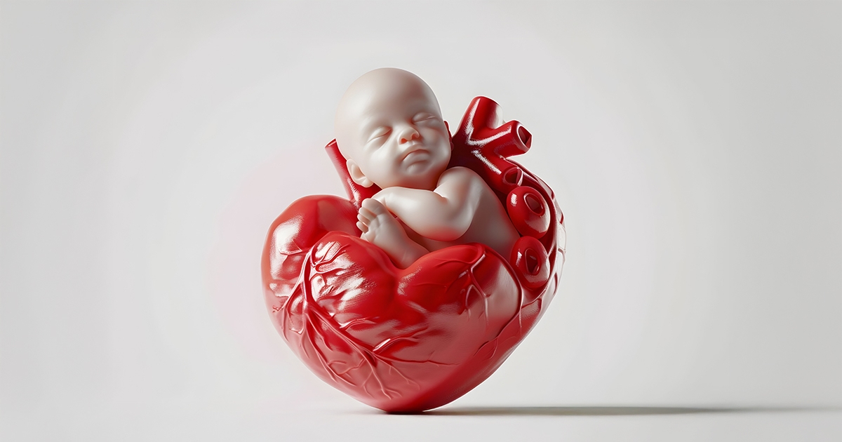
Congenital Heart Diseases in the Womb: Causes, Diagnosis, and Management
Fetal congenital heart defects represents the most common group of fetal anomalies, affecting approximately 1 in every 100 live births. It accounts for nearly 15% of neonatal deaths (within the first 30 days of life).
Understanding the causes, diagnostic methods, and latest treatment options is essential for early detection and effective management.
Who Is at Risk?
Fetal congenital heart defects, Interestingly, about 90% of congenital heart defects occur in pregnancies that are otherwise considered low risk. However, identifying mothers in the high-risk group remains crucial, as the likelihood of CHD is significantly higher among them.
Risk Factors
The risk of congenital heart disease increases under the following conditions:
- Presence of congenital heart disease in one or both parents
- Previous pregnancy loss due to a heart defect
- Maternal use of certain medications during pregnancy
- Early exposure to radiation (CT or tomography)
- Intrauterine infections during pregnancy
- Conception via in vitro fertilization (IVF)
- Chromosomal abnormalities in the fetus
In high-risk pregnancies, the likelihood of CHD ranges from 2% to 5%, meaning that 2–5 out of every 100 high-risk fetuses may develop a cardiac anomaly.
Diagnosis
Fetal heart abnormalities can be detected using the following imaging methods:
- Ultrasound (USG)
- Fetal echocardiography
- Color Doppler imaging
Fetal echocardiography is typically performed between 20–24 weeks of gestation. Detection rates vary from 10% to 80% depending on the expertise of the center, with top institutions achieving 70–80% diagnostic accuracy.
Detection Accuracy by Technique
- Four-chamber view: 20–40%
- Great vessels view: 50–60%
- Three-vessel view with color Doppler: up to 80%
Comprehensive ultrasound should always include detailed evaluation of the heart, brain, kidneys, gastrointestinal tract, and skeletal system, ideally in collaboration with pediatric cardiology and fetal radiology specialists.
When a Cardiac Anomaly Is Detected
Once a congenital heart defect is diagnosed, pregnancy and delivery management are individualized.
In cases where postnatal surgical correction is not compatible with life, pregnancy termination may be considered after thorough counseling.
Babies diagnosed with CHD should be delivered in well-equipped tertiary centers with:
- Perinatology and high-risk pregnancy specialists
- Neonatal intensive care units (NICU)
- Pediatric cardiologists and advanced monitoring equipment
If no chromosomal or structural abnormalities are detected, ongoing fetal surveillance is required to assess for the onset of heart failure.
When Should Fetal Cardiac Screening Be Done?
Recent studies suggest that cardiac assessment can be performed earlier than 20–24 weeks.
Some congenital heart defects can be detected as early as 14–16 weeks of gestation.
Conditions such as maternal infections, diabetes, epilepsy, radiation exposure, or medication use increase the risk, so even if a standard ultrasound appears normal, a fetal echocardiography and pediatric cardiology consultation are strongly recommended.
Is Nuchal Translucency Screening Only for Down Syndrome?
During the 11–14th weeks of pregnancy, if the nuchal translucency (NT) measurement exceeds 3.5 mm, amniocentesis is advised.
Even if chromosomal abnormalities are not found, an increased NT is associated with up to a 6% risk of congenital heart disease — thus, fetal echocardiography is essential in such cases.
Fetal Intervention: What Can Be Done Before Birth?
One of the most significant prenatal cardiac complications is fetal arrhythmia.
If the fetal heart rate rises between 200–450 beats per minute, immediate intervention is required.
If no structural defect is present, antiarrhythmic medications given to the mother can cross the placenta and restore the baby’s normal rhythm.
Treatment Options and Advances
When the fetal heart beats too rapidly, the heart cannot pump enough blood, leading to heart failure. In these cases:
- Medications can be administered directly via the umbilical vein.
- The goal is to stabilize the fetal heart rate and improve circulation.
In recent years, intrauterine balloon catheterization has been explored as a treatment for aortic or pulmonary valve obstructions, though this remains limited to a few highly specialized centers worldwide.
In Summary
Congenital heart disease is one of the most critical conditions that can be identified during fetal life.
With early diagnosis through fetal echocardiography, careful risk assessment, and multidisciplinary management, many affected babies can survive and thrive after timely intervention.