Prof. Dr. Arda Lembet » Imaging of the Female Heart: Diagnostic Methods and Significance
Imaging the female heart is critical because cardiovascular disease remains the leading cause of death among women worldwide. Despite this, heart disease in women continues to be one of the most frequently underdiagnosed and misinterpreted conditions in clinical practice.
This is not coincidental. Women develop heart disease through biological mechanisms that differ from those of men. When screening and diagnostic strategies fail to account for these differences, they often create a false sense of reassurance and lead to delayed diagnosis.
To accurately assess the female heart, imaging strategies must be reconsidered and redesigned in alignment with female cardiovascular biology.
In women, heart disease often does not follow the classic model of large coronary artery obstruction. Instead, conditions such as small vessel disease (microvascular dysfunction), endothelial dysfunction, and diffuse myocardial ischemia are more prominent.
For this reason, not all diagnostic tests provide the same value when evaluating cardiovascular health in women. Selecting the appropriate imaging modality is critical to achieving an accurate diagnosis.
This article presents a structured overview of cardiovascular imaging—from basic tests to advanced technologies—highlighting which tests are informative in which situations.
Electrocardiogram (ECG) An ECG measures the heart’s electrical activity and is typically the first diagnostic test performed. It is useful for detecting arrhythmias, signs of prior myocardial infarction, and acute ischemic changes. However, in women, significant heart disease may exist despite a normal ECG. This is particularly true in conditions involving the small coronary vessels, where the ECG’s sensitivity is limited. Thus, while ECG is a valuable starting point, it is not sufficient on its own to exclude heart disease in women.
Transthoracic Echocardiography (ECHO) Echocardiography is an ultrasound-based examination that assesses cardiac structure and function. It provides important information on ventricular performance, valvular disease, cardiomyopathies, and pulmonary pressures. However, ECHO does not directly visualize the coronary arteries and often appears normal in early-stage disease or in conditions affecting the small vessels.
Exercise Stress Testing (Treadmill or Stress ECG) Exercise stress testing evaluates the heart’s response to physical exertion. In women, however, hormonal influences, baseline ECG variations, and intrinsic test limitations contribute to higher rates of false-positive and false-negative results. The test is particularly inadequate for detecting microvascular ischemia and should not be relied upon as a standalone diagnostic tool.
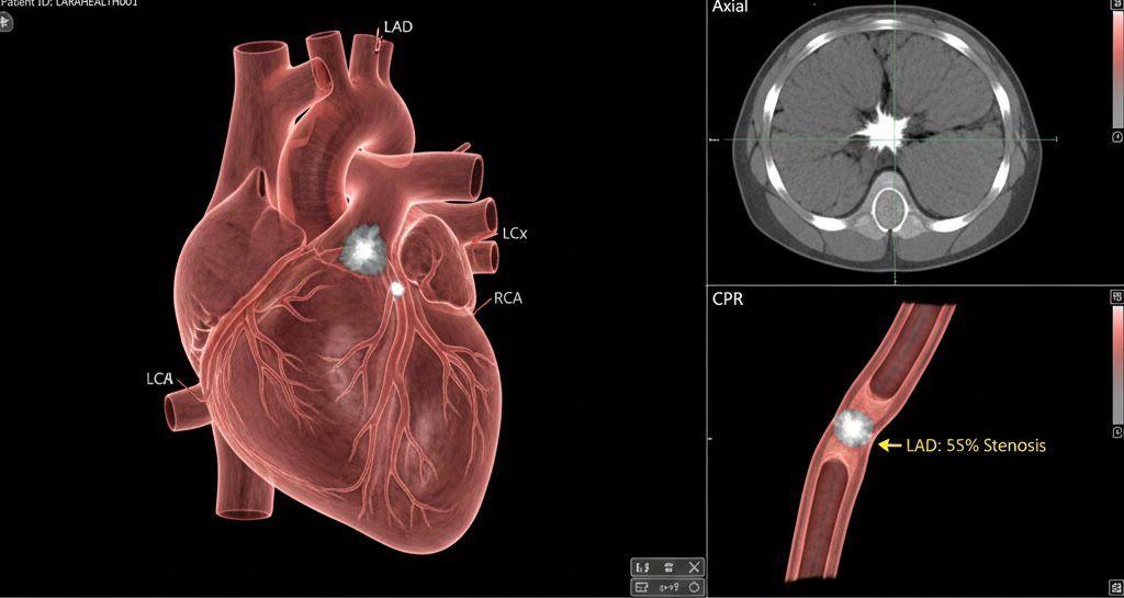
Coronary Angiography Coronary angiography is an invasive procedure that directly visualizes the major coronary arteries. It is essential for identifying significant large-vessel obstructions and guiding decisions regarding stent placement.
However, in women—especially in cases of INOCA (Ischemia with No Obstructive Coronary Arteries)—angiography may appear “normal.” This does not indicate the absence of disease and represents one of the key reasons why heart disease in women is frequently overlooked.
Coronary Artery Calcium Score (CAC) The coronary calcium score quantifies calcified plaque burden in the coronary arteries and is useful for detecting subclinical atherosclerosis before symptoms appear. It is particularly valuable in the perimenopausal and postmenopausal periods for predicting future cardiovascular risk.
Coronary CT Angiography (CCTA) CCTA provides detailed, noninvasive visualization of the coronary arteries. It can assess both calcified and non-calcified plaques. Its limitations include inability to directly assess microvascular function and exposure to radiation. Therefore, it is not suitable as a universal screening test.
Next-Generation Technology: Photon-Counting CCTA Photon-counting CCTA offers enhanced image resolution and improved visualization of smaller vessels. Despite these advances, even this technology cannot directly assess microvascular or endothelial dysfunction. It is a powerful tool—but not a comprehensive solution for all forms of female heart disease.
Stress Cardiac MRI Stress cardiac MRI is among the most comprehensive tools for evaluating myocardial perfusion and tissue characteristics in women. It accurately detects microvascular ischemia, diffuse perfusion abnormalities, myocardial fibrosis, and inflammation. Its lack of radiation exposure and strong performance in diagnosing INOCA make it one of the most valuable advanced diagnostic tests for symptomatic women.
Cardiac PET Cardiac PET quantitatively measures myocardial blood flow and is the most sensitive method for objectively assessing microvascular function. However, due to radiation exposure and higher cost, it is generally reserved for complex cases.
In asymptomatic women, evaluation should begin with ECG and echocardiography, followed by coronary calcium scoring to refine risk assessment. In symptomatic women, reliance on exercise stress testing alone should be avoided. Stress cardiac MRI should be prioritized.
If you are concerned about your cardiovascular health, you can contact our expert team. Importantly, “normal” ECG, ECHO, or angiography findings should not be interpreted as definitive reassurance.
Heart disease in women is not merely a matter of arterial blockage. It is closely linked to endothelial health, microvascular integrity, and regulation of myocardial blood flow.
Choosing the right imaging modality can make the difference between false reassurance and missed diagnosis. A truly woman-centered cardiovascular approach requires moving beyond traditional paradigms and adopting diagnostic strategies that place female biology at the core.
If you are re-evaluating questions about your own heart health, asking the right questions at the right time is a crucial first step. Approaching cardiovascular health with a holistic perspective is often the most powerful tool for early awareness and prevention.