What Is Chorionic Villus Sampling (CVS)?
Chorionic Villus Sampling (CVS) is a prenatal diagnostic procedure performed in early pregnancy to obtain a small sample of placental tissue (chorionic villi) for genetic analysis.
It is typically recommended between 11–14 weeks of gestation, particularly if the first-trimester combined screening or early genetic screening indicates an increased risk of chromosomal abnormalities.
Because the placenta and fetus share the same genetic material, CVS is a reliable method for detecting chromosomal disorders and inherited genetic conditions.
CVS is usually advised based on the mother’s family history, screening test results, or specific genetic indications.
Unlike amniocentesis, it does not provide information about neural tube defects such as spina bifida. Therefore, patients who undergo CVS are recommended to have a maternal serum test for neural tube defects between 16–18 weeks of pregnancy.
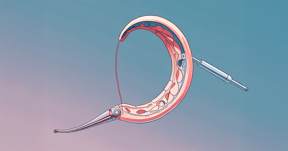
Procedure
CVS is recommended in cases such as:
- Pregnancies with a high risk of chromosomal anomalies
- Families with a known history of genetic disorders
It is generally performed between 10–12 weeks of gestation using either a catheter (thin tube) or a fine needle inserted through the abdomen.
Two main approaches are used:
- Transcervical Approach:
- A catheter is gently passed through the cervix under continuous ultrasound guidance.
- The catheter is directed toward the placenta, and a small sample of placental tissue is collected using gentle suction.
- Transabdominal Approach:
- A thin needle is inserted through the mother’s abdominal wall into the uterus to collect a small placental sample.
During the procedure, mild cramping sensations may occur.
The collected tissue is sent to a genetic laboratory for analysis, and results are typically available within 10–14 days.
In twin or multiple pregnancies, a separate sample is taken from each placenta.
Advantages and Risks
Advantages
- Enables early diagnosis of chromosomal or genetic abnormalities.
- If results are abnormal, early intervention or pregnancy management decisions can be made.
- If results are normal, it provides early reassurance for the expectant parents.
- Compared to amniocentesis, it allows for earlier detection in pregnancy.
Risks
- Miscarriage: The risk of pregnancy loss after CVS is similar to that of amniocentesis.
- Infection: As an invasive procedure, CVS carries a slight infection risk, especially if a vaginal infection is present. For this reason, a pelvic examination and culture are performed beforehand, and antibiotics may be prescribed if necessary.
- Limb anomalies: Procedures performed before 10 weeks of gestation have been associated with limb abnormalities in the newborn; therefore, CVS is not performed before the 10th week.
CVS, when performed by experienced specialists under ultrasound guidance, is a safe and precise diagnostic option that allows for early insight into your baby’s genetic health.