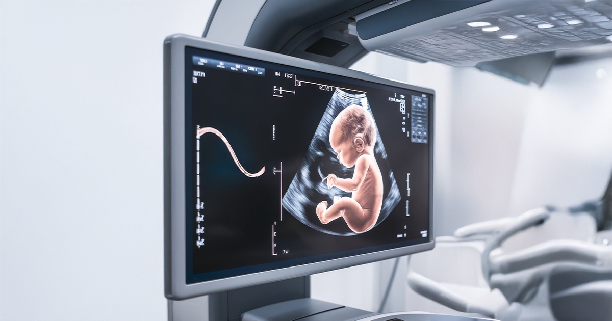
Which Methods Are Used to Detect Brain Anomalies in the Fetus?
At our clinic, between the 18th and 23rd weeks of pregnancy, a detailed ultrasound examination is performed to evaluate the fetal brain structure and to screen for both major and minor anomalies.
In cases where such anomalies are detected on ultrasound, a fetal MRI can be performed by our experienced team to obtain much more comprehensive and detailed information about fetal brain development.
Case Example
In the first pregnancy of our 29-year-old patient, an ultrasound examination performed at 23 weeks of gestation revealed that the fetal head circumference was below the normal range.
The patient was screened for TORCH infections—a group of infections that can affect the fetus during pregnancy. In addition, amniocentesis was performed, and the fetal karyotype was analyzed from the amniotic fluid sample.
A fetal MRI conducted at 26 weeks of gestation showed that the fetal head circumference remained slightly below average.
When the MRI was repeated at 31 weeks, flattening of the cerebral gyri and a marked decrease in head circumference were observed.
Based on the confirmatory findings obtained from the fetal MRI, termination of pregnancy was planned at 32 weeks of gestation due to the risk of abnormal fetal brain development.