
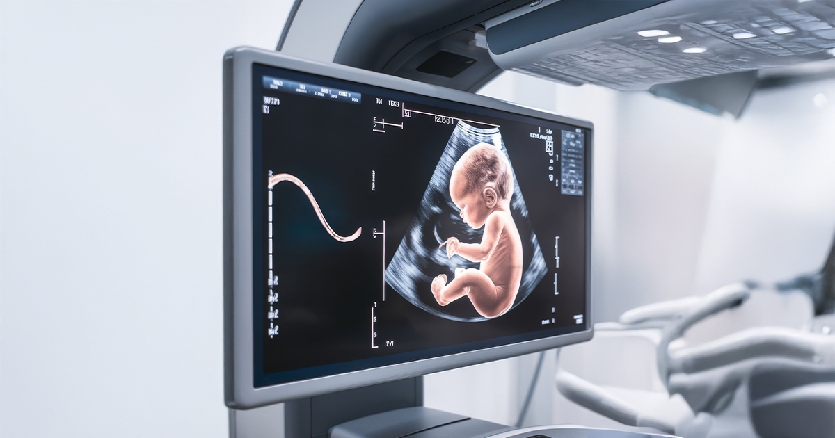
Fetal neurosonography involves specialized ultrasound imaging designed to map the fetal brain between 18 and 23 weeks of gestation. This examination allows for the identification of both major structural anomalies and minor developmental deviations.
In instances where ultrasound findings are ambiguous or reveal potential anomalies, fetal MRI is utilized to provide high-resolution, comprehensive data regarding brain structure and architecture.
Clinical Case: Monitoring Fetal Brain Development
A 29-year-old patient presented at 23 weeks of gestation with an ultrasound indicating a head circumference below the standard growth percentile. The clinical management followed a rigorous multi-step diagnostic protocol:
Infectious Screening: The patient was tested for TORCH infections (Toxoplasmosis, Other agents, Rubella, Cytomegalovirus, and Herpes simplex) to exclude congenital infections as the underlying cause.
Genetic Assessment: Amniocentesis was performed to obtain a fetal karyotype, ensuring chromosomal stability.
Serial Imaging:
26 Weeks: A fetal MRI confirmed that the head circumference remained below the expected average.
31 Weeks: A follow-up MRI revealed significant markers of abnormal development, specifically the flattening of the cerebral gyri (lissencephaly-like appearance) and a progressive decrease in head circumference relative to gestational age.
Management Decision: Given the confirmed MRI findings suggesting severe neurological impairment, a pregnancy termination was planned at 32 weeks of gestation.
This case highlights the integration of neurosonography, genetic testing, and serial MRI imaging in the precise diagnosis and management of severe fetal brain pathologies.